Hmm have a needeled syringe ?
Tip the bottleneck 45 degrees an see if they accumulate then suck it up
See wat it is water??
2 Likes
I added more olive oil, warmed and stirred on a hot plate. Droplets are now gone.
I may try to do a bigger batch and use the needle Trick to try to grab a sample.
2 Likes
They will come back most likely if given time to Setlle and seperate
2 Likes
Did the olive oil get cold? Often when olive oil gets cold enough, the longer chain triglycerides start to solidify and seperate. You can see this by keeping olive oil in the fridge or living in Canada in the winter ![]()
3 Likes
Strike that it appears to be of the same fluorescence as cannabis extract, but apparently the “sauce” part more then the thc.
2 Likes
It didn’t get below room temp, around 70F.
It also didn’t happen to the straight olive oil that was right next to it, only the cannabis infused olive oil. I’m going to pop it into the fridge/freezer to see what it looks like when it is cooled.
It’s been about 8 hour and there hasn’t been any separation since I added more olive oil.
2 Likes
Interesting either way
1 Like
Someone out there should get their hands on some of that fake shatter going around and see if it glows any different than authenic samples. It would be pretty cool if there was any observable differences and would be a great addition to the isopropyl alcohol test.
(just a pic of some ethanol shatter)
6 Likes
Woah, how did I miss this thread! I was just thinking about this topic recently.
In fact, @phenethylamine1, I did a brief TLC experiment like you do and had some interesting observations myself. From playing around with the UV light (long wave) on distillate in jars or short path, I had always thought THC was kind of fluorescent. I now do not think pure THC is particularly fluorescent.
I was doing experiments on acidic water->basic water/heptane LLE on winterized CO2 crude I dissolved in heptane. I was trying to see if it was possible to use chromatography on the crude to separate out THC , or at least clean up the crude a lot. I ran the crude on TLC and of course got some really lovely isolation of very pretty colors. I then compared this to ~80% distillate on the same TLC. Surprisingly, the THC spot didn’t really fluoresce at all under the long wave, it was more of a dark spot on the TLC.
Also, to my surprise, while the LLE on the crude didn’t seem to significantly improve potency or color of the final distillate, it DID greatly dampen the fluorescence of the distillate product! I can’t seem to find a the picture I took, but I put two test tubes side by side of distillate made from the same batch of crude, one with LLE treatment and one without, and there was a noticeable difference.
Also, I just help up a UV lamp to some old jars of distillate I have and it appears that the top layer, where air exposure is the greatest, is fluorescent green, the rest is only a very dull fluorescence.
This leads me to believe that THC oxidation products -or some trace contaminant that oxidizes but isn’t THC- is fluorescent, but pure THC isn’t. I like your test with the decarbed THCA, but doing it in a solvent under inert conditions might be a better test to see if THC is actually fluorescent.
In conclusion… I dunno
5 Likes
This is very interesting information. Thank you for sharing. I agree with you for sure that the glow may be in part from degradation. My decarb test on the capillary tube was done in atmospheric oxygen with a butane lighter (uncontrolled temp). When I decarb in a controlled setting, the glow appears, but way way less intense.
Your LLE/TLC experiment sounds interesting. I’m going to have to think about it for awhile.
Yeah, total confirmation on glow amplification from degradation.
1 Like
I would love to see fluorescence observations for decarbed THC in a pure nitrogen or argon environment!
Also, do you (or does anyone reading this thread) have access to interlibrary loan? I am trying to pull this paper, titled “Fluorescence of cannabinoids” from LibGen but it’s not finding it:
https://onlinelibrary.wiley.com/doi/abs/10.1111/j.2042-7158.1975.tb09424.x
1 Like
https://moscow.sci-hub.tw/5100/5da92056401ccbd1cb5b6c9e92a7bd9e/dionyssiou-asteriou1975.pdf Try this
3 Likes
Thank you my friend!
1 Like
What kind of UV light do you use? And what do the UV lights actually show? I am getting conflicting answers and am just curious where you got your information from.
A simple black light
did you start at the top?
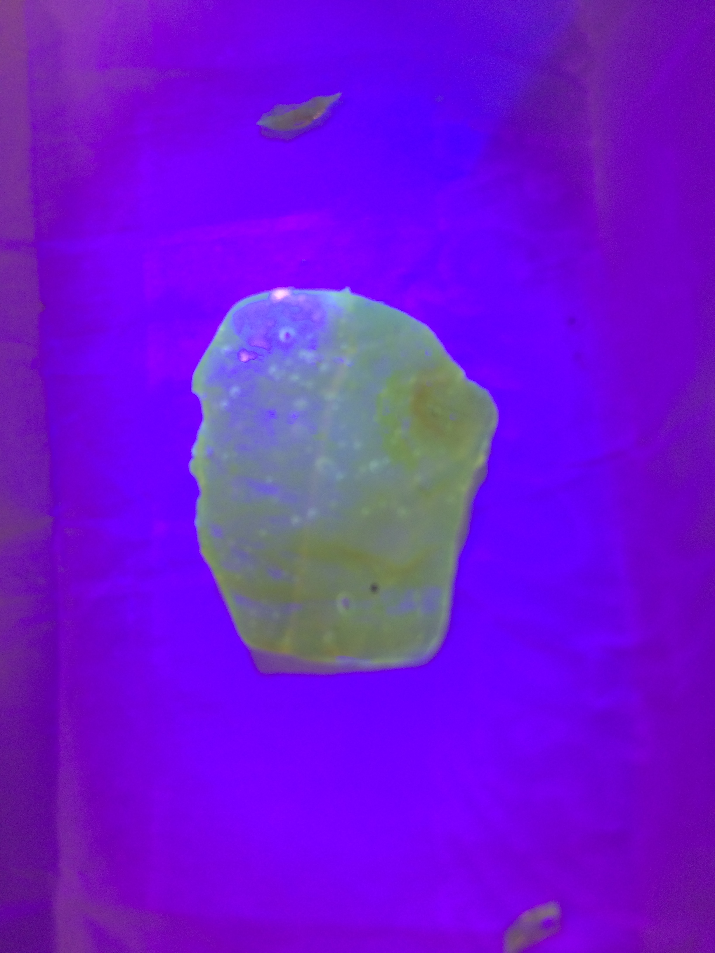
Ultraviolet Fluorescence of Cannabis - Observations
you’re repeating the first question asked…
which was answered with link by the OP in post three.
these are first hand reports. with pictures. which literally show what UV shows…
where are you getting your conflicting reports from? (links please).
Edit: if you want to dig into the whys and wherefores, you should visit the data dump and look for the extinction coefficients of the various cannabinoids. Or check up thread to make sure so me kind soul hasn’t linked those already.
6 Likes
Great thread, I hopefully will have something to contribute in a few weeks as i just picked up a dual wavelength wand normally used for TLC but I’ve been doing some normal phase chromatography and just waiting on my c-18 for reverse phase. I wonder what I will see in my columns ![]()
![]()
5 Likes
I wanted to come back to this topic,
I currently have access to a fluorimeter in my lab, it basically scans different wavelengths. Would this be able to identify different cannabinoids in a mixture or an isolate given each has a slightly different emission spectrum?
Based on my observations thc is visible at 800nm whereas minors and cbd are not. This is extremely useful for anyone who has uv detection on their fractionation systems. As long as it scans from 200-800nm
4 Likes
