I’ve made observations of UV fluorescence during cannabis processing that I’d like to share.
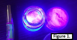
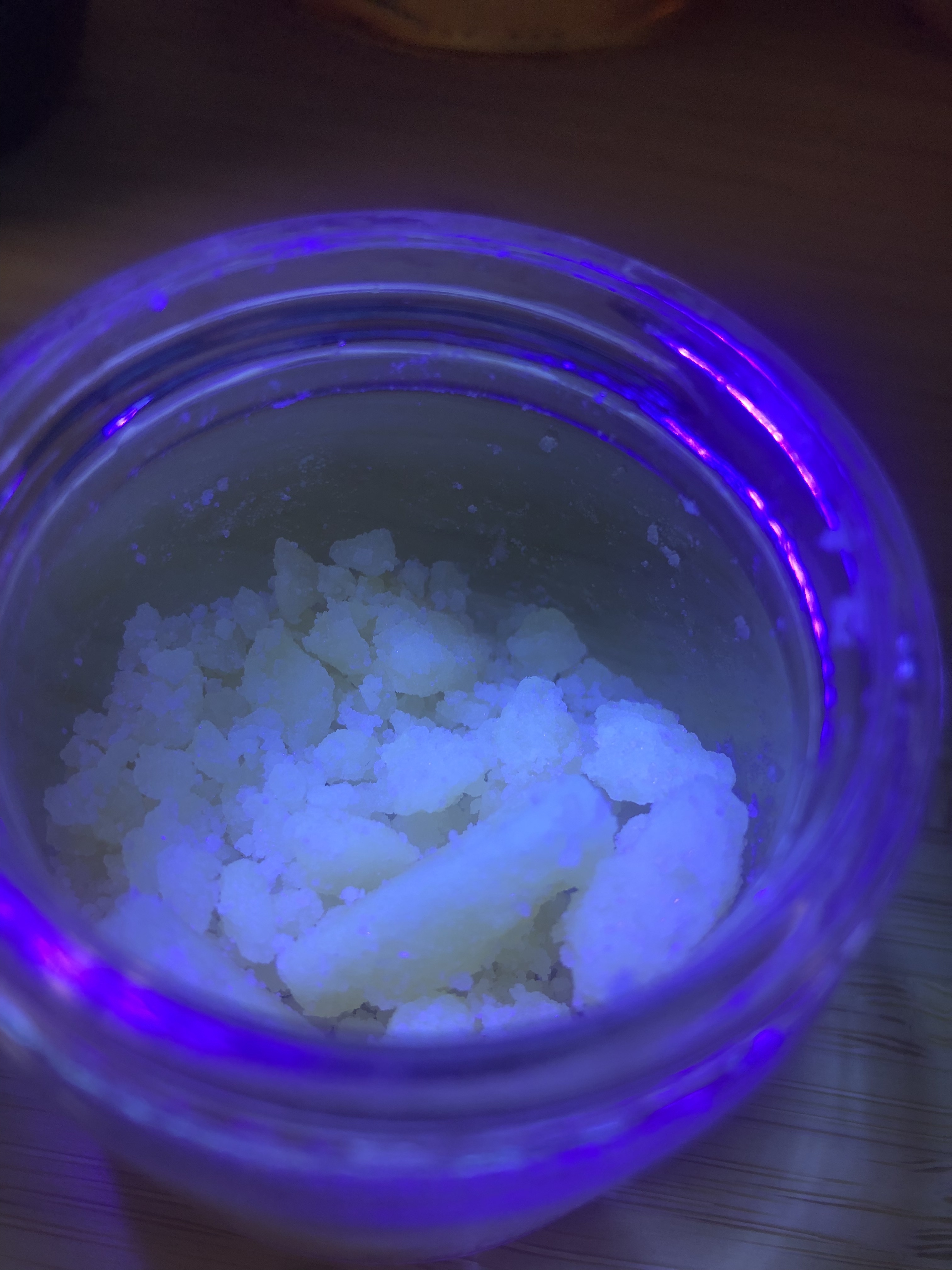
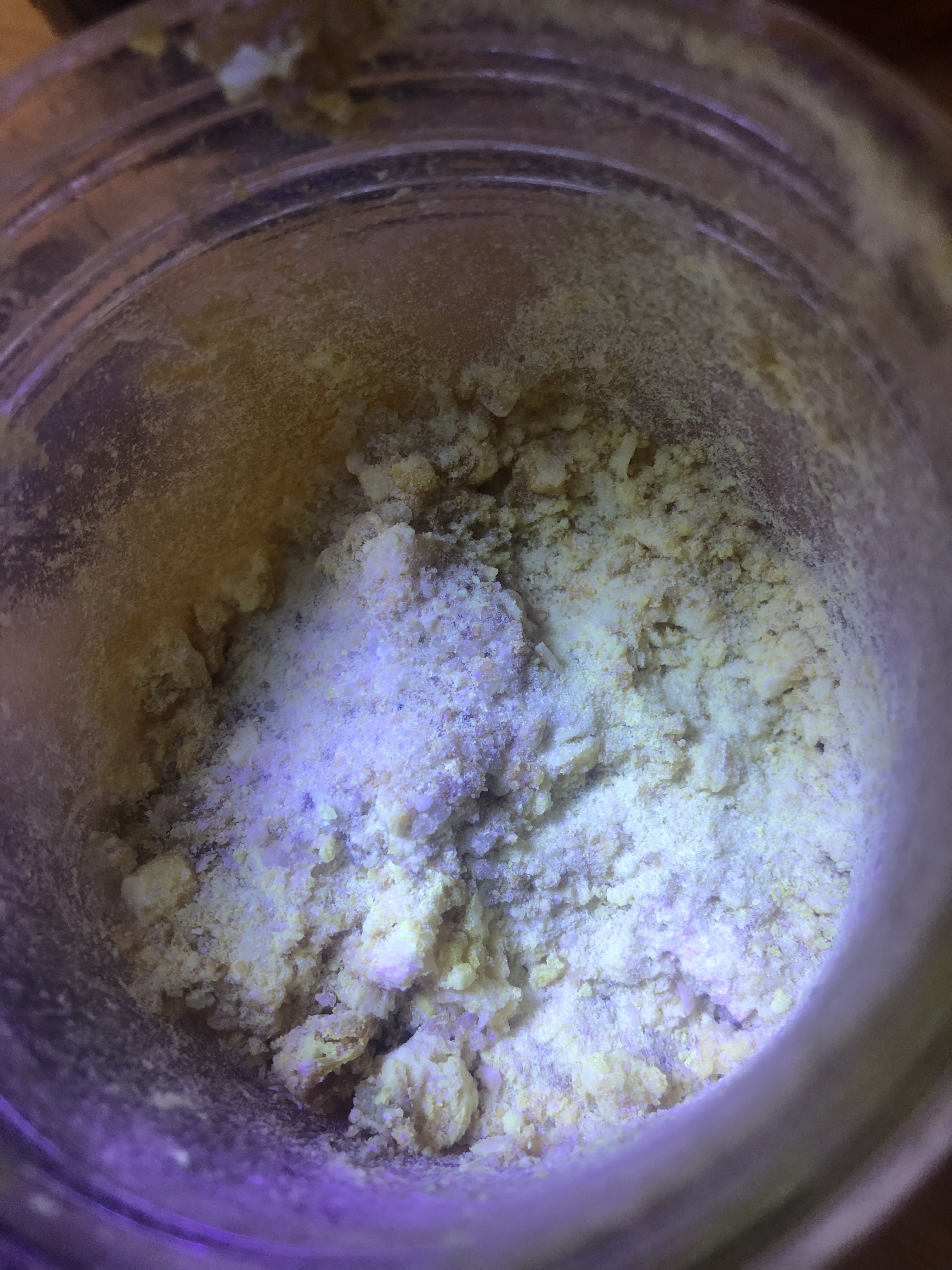
(Fig 1) as shown from left to right is a 78% total cannabinoid distillate, 98% THCA isolate, and 99.8% CBD isolate. The distillate has a green glow and the isolates have none.
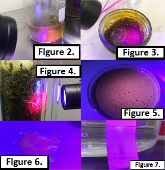
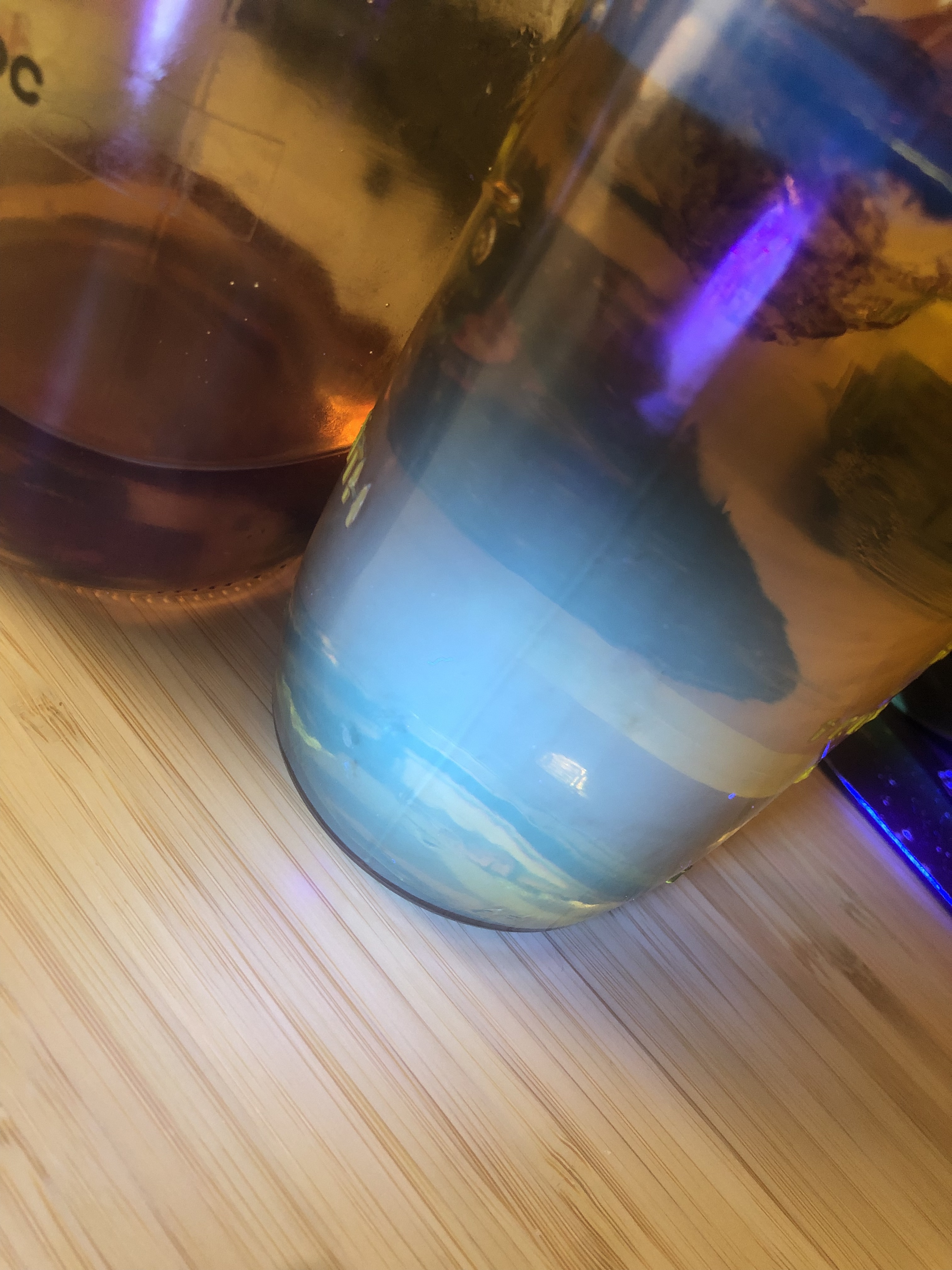
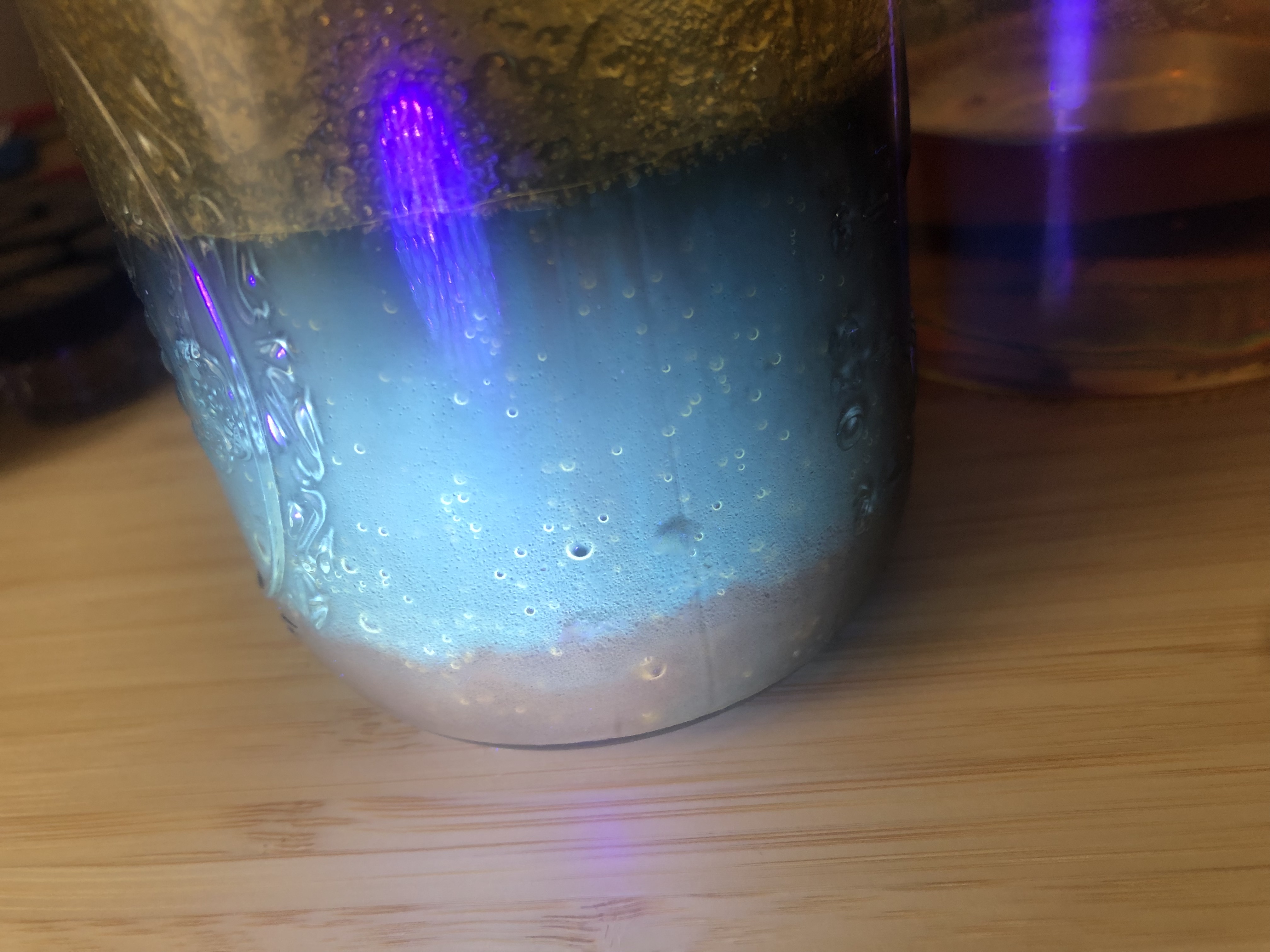
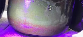
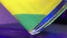
(Fig 2) is an ethanol extraction at -78C with 20 micron inline filtration approximately 190 proof. (Fig 3) is crude hydrocarbon/CO2 extract. (Fig 4) is a soxhlet extraction with ethanol. When dissolved is 99% isopropyl alcohol and boiled, the red fluorescence precipitates and floats on the surface of a now brown fluorescent material or mixture in (Fig 5). If extractions with most polar solvents are done below -80C the red material is captured with cellulose filters in Figure 6. On a cellulose TLC strip using 70% ISO or 66% Ethanol mobile phase, crude extracts of all types separate into a high retention red fluorescent group and low retention green grouping in (Fig 7).
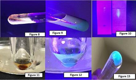
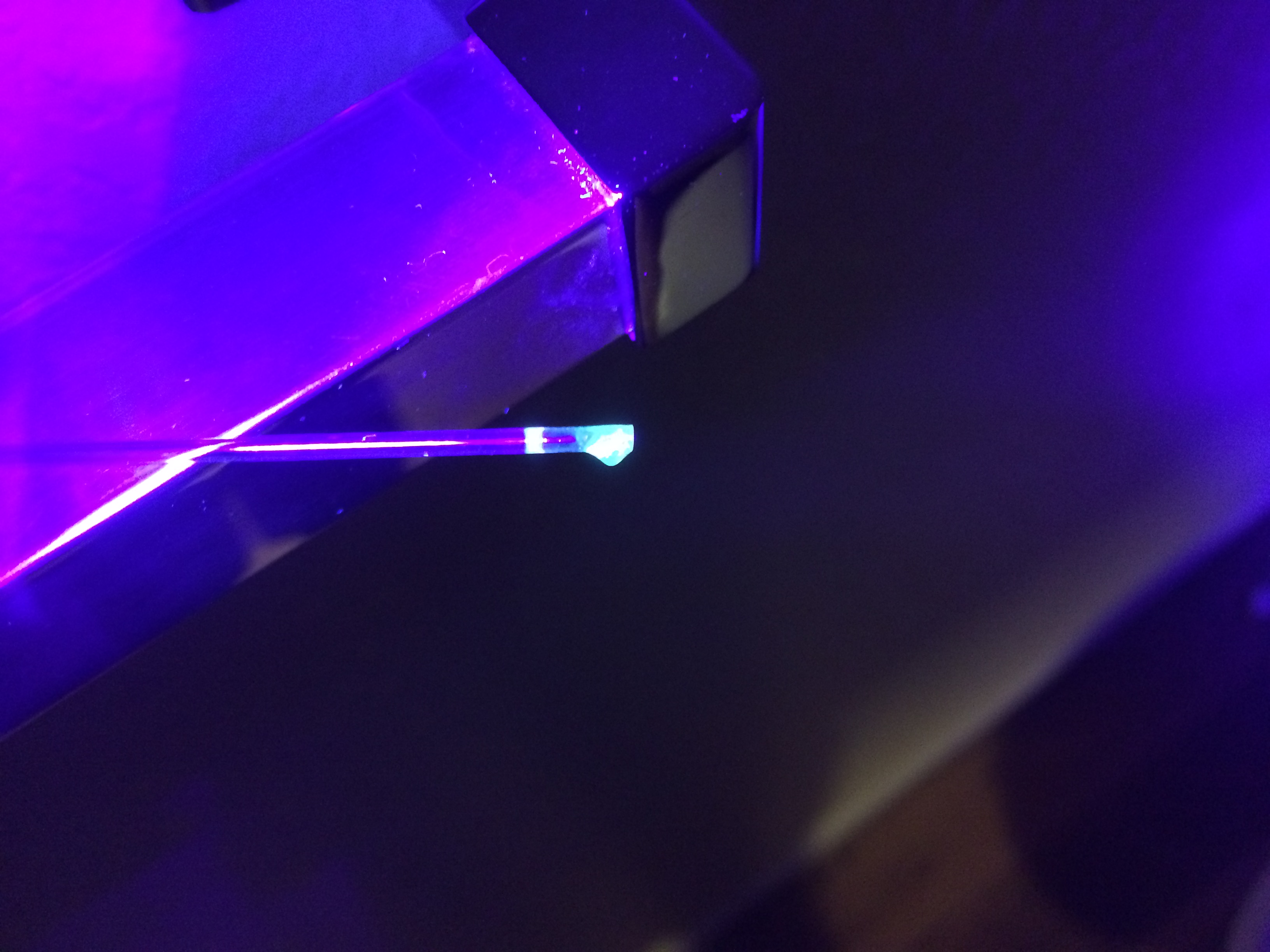
(Fig 8) presents a crude extract made from room temperature 190 proof ethanol extraction as a brown/red glow. The solvent was evaporated and extract redissolved into 99% ISO. The ISO/Extract solution is washed with naptha and the brown/red glow is transferred to the organic layer, and the green glow is left behind in the 99% ISO in (Fig 9). When the two fractions are examined under TLC using 70% ISO mobile phase on cellulose, the organic layer (left strip) has no mobility and trace green glow while the ISO layer (strip right) has almost complete mobility up the strip in (Fig 10). The ISO layer is evaporated in a test tube and when the oil separates, fluorescence is transferred to the top layer and the bulk of the oil sinks (Fig 11) (Fig 12). The lower layer is pipeted into a new test tube and evaporated yielding a bright blue flurecent oil (Fig 13).
In (Fig 14) the test tube on the left is plant material boiled in distilled water exhibiting blue/green fluorescence and the test tube on the right is a plant material boiled in a non-polar solvent. (Fig 15) is store bought extract of an unknown method exhibiting blue glow. (Fig 16) Blue fluorescent material gathers on the head on wiped film during a repass of 78% distillate. (Fig 17) Repassed oil with blue flurecent material removed and overall fluorescence decreased proves to contain more cannabinoids by weight.
What type of uv light is used to lighten up the samples ?
Nice tread by the way
Years back it was metioned somewhere
And i always have one close
But lately i haven t done any thing with it
Since you started i might as well
I always had the feeling a good heads ,mains tails moment could be achieved with uv lighting on the condensor
But until i noticed diffre nt biomass difrent light show
I’ve used quite a few different light’s and they all seem to produce very similar if not almost identical glows. Here is the kit I have now, I like it because it comes with eye protection:
I’ve noticed that on SPD the head fraction glows quite strongly blue and changes to green when the main delta 9 fraction comes along. Isomerization also makes changes in the glow too. I’d love to see what other peoples observations are with different extraction methods and different samples. Like what does pure CBC, D10, CBN, CBG or other cannabinoids glow like?
This is awesome. Thank you for your share. I’m curious if somebody can get there hands on any of the fake shatter that can hardly break and burns weird. I wonder if this method could be used to find fake concentrations?
I’d bet you $1 the red color earlier in the thread is chlorophyll fluorescence. You can even visualize this with a strong white light source aimed at the side of an olive oil bottle (assuming the glass is clear). I published a series of papers in the late 80s, early 90s on video techniques to estimate photosynthetic electron transport via fluorescence analysis, so I’ve seen that color a lot!
Nice layering on the bottom jar. I’ve had precipitation happen like that by changing the proof of ethanol solutions. The first to precipitate out is red and if you precipitate the green again you get yellow top, blue green bottom. The yellow proved to contain quite a bit of oil that tasted like pineapple heaven.
Balachandran, S., Osmond, C.B., Daley, P.F. 1994. Diagnosis of the earliest strain-specific interactions between tobacco mosaic virus and chloroplasts of tobacco leaves in vivo by means of chlorophyll fluorescence imaging. Plant Physiol. 104: 1058-1065.
Daley, P.F. 1995. Chlorophyll fluorescence analysis and imaging in plant stress and disease. Can. J. Plant Path. 17: 167-173.
Daley, P.F., Raschke, K., Ball, J.T., Berry, J.A. 1989. Topography of photosynthetic activity of leaves obtained from video images of chlorophyll fluorescence. Plant Physiology 90: 1233-1238.
Osmond, C.B., Daley, P.F., Badger, M.R., Lüttge, U. 1998. Chlorophyll fluorescence quenching during photosynthetic induction in leaves of Abutilon striatum Dicks. Infected with Abutilon Mosaic Virus, observed with a field-portable imaging system. Bot. Acta 111: 390-397.
We reduced this to a field-portable setup for plant disease studies, but it was early 90s, so there were some tech hurdles that have disappeared since…science marches on!
Found something interesting. I decarboxylated the THCa in a capillary tube and it took on a green glow, then roasted the pooled extract over a butane cigarette lighter and it takes on blue glow just like isomerized oil or short path heads.